

 There is a study which must be done within the first three months of pregnancy, between the 11th and the 14th week of gestation, ideally at the 12th week, in which currently its been the chosen method of screening for detection.
It has as general objective to detect in early way if a baby is in risk of developing any kind of syndrome. Among the most frequent are Down syndrome, even though at this age is also reach to detect another structural abnormalities, such brain defects, urinary tract, spine, facial defects, walls of abdomen defects on the baby, and suspicion of some kind of heart abnormalities.
 As an advantage, its also an opportune moment for predicting the risk of adverse perinatal outcomes such as early pre-eclampsia before 34 weeks or early growth restriction by doppler measurement of uterine arteries
Who are candidates for this type of study?
The ideal would be to perform it on all pregnant women, but this risk increase after 35 years old.
What parameters should be measured in this study?
Basically the sign that is looking for is the nuchal translucency that corresponds to a space filled with liquid in the nape of the baby, which its identified increased on Down Syndrome, therefore, a detection strategy is the measurement of this ultrasound sign.
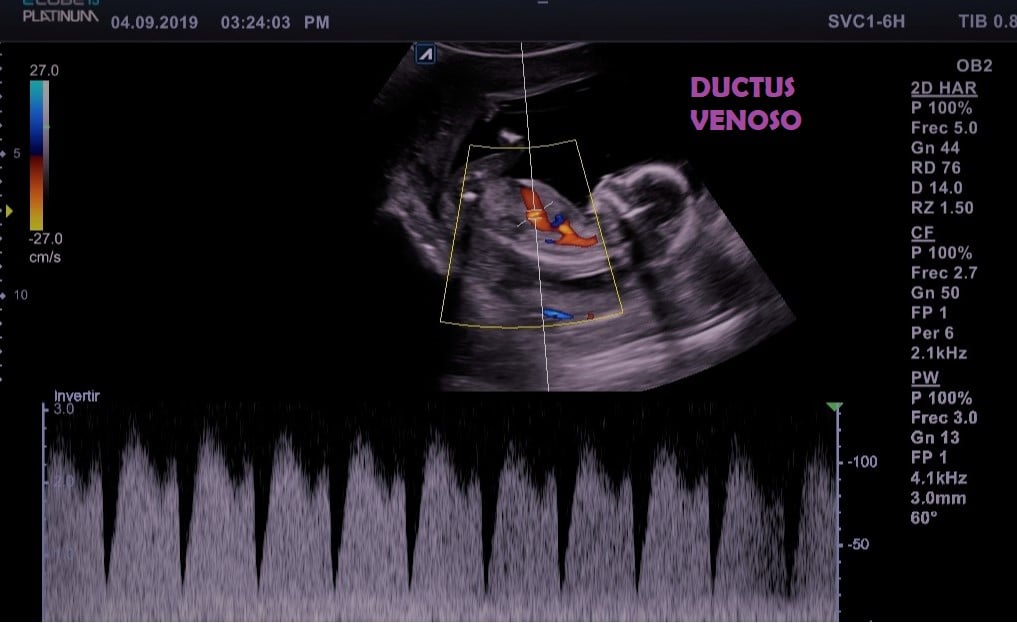
Also exist other markers such as the nasal bone, which is absent or smaller and has been seen in babies at risk of Down Syndrome . The ductus venosus which is a very important venous vessel since it carries blood oxygenated directly to the inferior vena cava, since it is found with a reverse spectrum in babies at risk of Down syndrome and also in babies who develop any abnormality of the heart, another sign also described is the regurgitation of the tricuspid heart valve that occurs in babies at risk of Down syndrome
Since a decade, specialists and pioners in this subject, such as Dr. Nikolaides have suggested making and investment in terms of the pyramid of prenatal care, since traditionally the structural evaluation of babies were made after 19 or 20th weeks of gestation, and as a progress has been made a development of this knowledge, the improvement in image quality with higher resolution ultrasound its currently feasible to carry out an early evaluation for certain anomalies such as diseases of brain, holoprosencephaly, anencephaly, facial cleft lip, thoracic suspicion of diaphragmatic hernia, abdominal wall defects such as gastroschisis, omphalocele herniation of the contents of the abdomen, urinary tract disorders such as enlarged bladder, or megabladder.

This type of study, can be combined with biomarkers in maternal blood, pus a series of maternal characteristics that when entering all the patient information in a software designed by the Fetal Medicine Foundation performs a risk calculation with a detection rate of more than 90%, with a low false positive rate of less than 5 %.
In the absence of markers in maternal blood, the detection rate certainly decreases, but the test performance considering nuchal translucency plus maternal characteristics is close to 80% already described in well validated studies.
In order to carry out these studies, training an a certification is required to guarantee being able to carry out the measurements, granting a license to perform the calculations.
Its important to mention that this test only detect risks, and when detecting a fetus with high risk, a diagnostic test and advice from a multidisciplinary team fetus should be offered.
Dr. Adrián Israel Ramírez Mendoza
Especialista en Materno Fetal